
 WHAT IS A MYELOGRAM?
A Myelogram is a diagnostic imaging procedure completed by a radiologist. Such imaging can help identify problems within the spine. If experiencing symptoms related to a spinal condition, the problem could be the nerves, the spinal cord, or the surrounding tissues. Pinpointing the location of the problem assists with diagnosis and coming up with effective treatment plans.
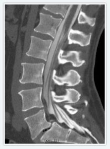
This procedure involves the use of contrast dye which is injected into the spinal column before the procedure. An X-ray is taken and the contrast dye allows the radiologist to see the spinal cord and other areas of the spine more clearly than a regular X-ray. Along with the spinal X-ray, your radiologist performs a CT scan during a myelogram. This is an imaging test that uses X-rays and a computer to create detailed images of the spine, and is much more detailed than a standard X-ray. These procedures allow medical professionals to examine the spine in detail. They look for deformities, weak areas, compression, and signs of trauma.
MYELOGRAPHY OR MYELOGRAM PROCEDURE?
Myelography and myelogram are the same test, so the words can be used interchangeably. While myelography has been used in the past as the best way to view the inner workings of the spine, these days MRI scans have overtaken traditional X-ray and CT scan myelography. Using an MRI has many advantages, such as lack of radiation, being non-invasive, and there is no need for intrathecal contrast material. Though the non MRI myelography will still be used in the case that the patient cannot have an MRI scan, such as due to a pacemaker or implants.
WHAT IS A CT SCAN?
The term CT is short for Computed Tomography. The basic premise of a CT scan is the computer assisting part, which compiles multiple X-rays from multiple angles to create a more complete picture of the spine or inner body. Spine pictures and images are captured from different angles and various areas so that the exact problem area can be diagnosed by medical professionals. This procedure allows more details and information to be seen than with a typical X-ray only.
SPINE X-RAY EXPLAINED
X-rays are a high energy electromagnetic radiation, and can be used to create images of inside a person’s body. How this works is due to different parts of the body absorbing different amounts of radiation, like the bones absorbing more X-rays than anything else so they show up white. When someone starts exhibiting possible symptoms of a spinal condition, an X-ray can be a useful tool. They may be struggling with balance or coordination due to spinal concerns. The images taken can show the severity of the problem and help the doctor determine the next steps. This can include an exercise plan and physical therapy, as well as medication if needed.
If a Myelogram shows serious problems or advanced deterioration in the spine, surgery may be necessary. The doctor can share the images taken during the procedure to explain why they feel this is the best course of treatment. Otherwise, the problem and the pain can continue to get worse with time.
 WHEN YOU MAY NEED A MYELOGRAM
Your doctor may send you for a Myelogram based on their initial assessment. Talk to them about any symptoms or concerns you may have. If they believe there could be an underlying spinal issue, the CT and spine X-ray will help them confirm the initial diagnosis. Some of the reasons you may be sent for such imaging include:
- Arthritis in the discs of the spine
- Bone diseases in the spine
- Bone spurs
- Cysts
- Herniated disc
- Infection or inflammation in the spinal area
- Spinal nerve damage
- Tumors in the brain or spinal region
GETTING READY FOR THE PROCEDURE
Make sure you share any details that could affect your imaging. Women who are pregnant or may be pregnant need to let your doctor know as imaging may not be a good idea during pregnancy. Let them know if you take any antibiotics or blood thinners. If you are ill or have an infection it is best to reschedule the Myelogram until you feel better. This will help ensure accurate results and safety during the scan.
Depending on the facility, you may be able to keep your own clothing on. Wear loose-fitting clothing that is easy for the radiologist to shift around if necessary. Other locations will ask you to change into a medical gown for the procedure. Stay well hydrated 48 hours before your procedure. Make sure you don’t eat anything in the 3-hour window before your procedure.
HOW IS A MYELOGRAM PERFORMED?
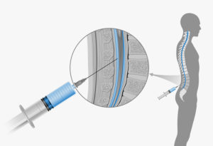
Many facilities require you have someone with you for emergency reasons and to drive you home. It is recommended not to drive for 24 hours after the imaging is conducted. When it is time for the procedure, you will lie on a bed on your side. Your spine and back will be cleaned with antibacterial soap. Local anesthesia will be administered.
![]()
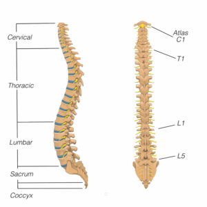
Once the area is numb, the puncture is made at the lumbar area and the contrast dye is injected into the spine. You may feel pressure when the needle goes into the spine but it shouldn’t hurt. You will need to remain as still as possible during the procedure. Then the X-ray and CT imaging scans are performed. The time involved is usually 30 minutes to an hour, but can vary from patient to patient.
AFTER THE PROCEDURE
You will need plenty of rest after the procedure. It’s important to drink lots of fluids afterwards. You may need to stay in the recovery area for a couple hours before they release you to go home. Make sure you let health care professionals know about any symptoms you have including nausea, pain, tingling, or headache.
The results of your imaging will be sent to your medical professional for them to review and evaluate. They can discuss the findings at your next scheduled appointment. With this information, they may schedule more tests or have enough to make an accurate diagnosis.
POTENTIAL MYELOGRAM RISKS
While most people have no troubles with a Myelogram, there are some potential risks to be aware of. Much of the risks or side effects are associated with the contrast dye being injected into the spine.
 CONTRAST DYE SIDE EFFECTS
Though rare, some individuals have an allergic reaction to the contrast dye used in the imaging. If you have ever had a bad reaction to any contrast make sure your medical professionals are well aware of it.
Fluid may leak from the puncture at the lumbar area where the contrast is inserted. This can cause headaches that last for a few days. A small amount of CSF leakage is common but if it is a large amount or continues for several days let your doctor know. It can also trigger severe headaches if the leak is a large amount. There may also be aches in the arms, legs, nausea and dizziness. But most patient experiences no side effects whatsoever, and if they do they subside within 24 hours.
Seek immediate medical attention if you develop a fever. There is a very rare risk of bleeding in the spinal canal when a Myelogram is conducted.
HOW LONG DOES IT TAKE FOR CONTRAST DYE TO LEAVE THE BODY?
Constrast dye should be completely eliminated from the body within 24 hours, but usually in as little as 3 hours.
It is best to have someone drive you after the procedure. Some patients experience numbness in the legs or their lower back. This can make it hard to walk out of the facility too. Let the radiologist know if you feel such symptoms. They can give you a place to rest until the numbness goes away or get you a wheelchair to be taken to the vehicle. You don’t want to risk a fall or other injury.